People have been using heart as a symbol for presenting love, mercy, emotion and passion since centuries. It represents our emotion and health. We have been taught to draw ♥ when we want to tell someone we love you since childhood. The majority of children draw themselves with their parents around it with smiles in their faces under sun shine in the middle of a garden. However, some other people complain because their heart has been recently broken due to a break up or because there is a medical problem.

In fact, the real heart is not simple as we thought. It is very complex to serve our life. It pumps blood to our body all the time without a break. Can you do it? Can you work 24/7 without taking rest? It can.
This organ consists of 4 chambers; 2 of them for receiving blood and the other 2 are for pumping blood. When we draw a heart, we should consider that we draw someone's heart who is looking at us; that means both right Atrium and Ventricle are on the left and both left Atrium and Ventricle are on the right side.
Lets see how we can draw a heart so easily by this awesome video:
For donation: http://www.handwrittentutorials.com/donate.php
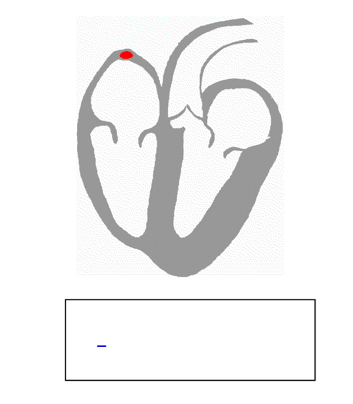
The real heart looks like this:

Blood circulation:
There are 2 types of blood circulation
1. Major circulation (systemic circulation): the blood is pumped from the heart to the body through Aorta and vice versa through both superior and inferior Vena Cava.
2. Minor circulation (Pulmonary circulation): the blood is pumped from the heart to the lungs through Pulmonary arteries and vice versa through Pulmonary veins.

Some important definitions:
1.Cardiac cycle: Sequence of events in 1 heartbeat. Blood is pumped through the entire cardiovascular system.
2.Systol: Contraction phase, usually refers to ventricular contraction.
3.Diastol: Relaxation phase, the atria and ventricles are filling. Lasts longer than systole.
4.Stroke volume (SV): Amount of blood ejected from either ventricle in a single contraction.
5.Starling’s Law of the Heart states that degree of cardiac muscle stretch can increase force of ejected blood. More blood filling the ventricles ↑ SV.
6.Cardiac output (CO): Amount of blood pumped through the cardiovascular system per min.
CO = SV X Heart rate (HR)
7.Depolarization: The electrical charge of a cell is altered by a shift of electrolytes on either side of the cell membrane. This change stimulates muscle fiber to contract.
8.Repolarization: Chemical pumps re-establish an internal negative charge as the cells return to their resting state.
The conduction system of the heart:
Several types of cardiac muscle fibers exist within the heart. It is often useful to classify these types broadly as either contractile or conductile.
CONTRACTILE cells are the cells of the working myocardium and constitute the bulk of the muscle cells that make up the atria and the ventricles. An action potential in any one of these cells leads to vigorous force development and/or mechanical shortening.
CONDUCTILE cells are specialized muscle cells that are involved with the initiation or propagation of action potentials rather than direct generation of force. In addition to the effects of the CNS on the heart, the heart can work on its own. The conducting system of the heart consists of cardiac muscle cells and conducting fibers (not nervous tissue) that are specialized for initiating impulses and conducting them rapidly through the heart. They initiate the normal cardiac cycle and coordinate the contractions of cardiac chambers. Both atria contract together, as do the ventricles, but atrial contraction occurs first. The conducting system provides the heart its automatic rhythmic beat. For the heart to pump efficiently and the systemic and pulmonary circulations to operate in synchrony, the events in the cardiac cycle must be coordinated.

This conduction can be summarized as the following:
From Sinoatrial node (60-100 times/minute, bpm) to Atrioventricular node (40-60 times/minute) to bundle of His to left and right bundles to Purkinje fibers (20-40 times/minute).


Types of pacemakers:
SA node is the natural (main) pacemaker of the heart.
AV node, bundle of His and Purkinje fibers are the latent pacemakers.
SA node is the natural (main) pacemaker of the heart.
AV node, bundle of His and Purkinje fibers are the latent pacemakers.
Cardiac action potential:


1.At membrane potential -60 mV, Na/K channels (funny channels) open to influx Na and efflux K [GRADUAL DEPOLARIZATION].
2.When membrane action potential reaches the threshold at -40 to -45 mV, T Ca channels open (T stands for Transit = FAST)
3. L Ca channels open (L stands for Long = SLOW) for Ca influx till the action potential reaches 0 mV [SPIKE or FAST DEPOLARIZATION].
4.At this point, K channels open to facilitate K efflux.
5.That makes the cytosol increasingly negative and creates the following phase of action potential [REPOLARIZATION].
6.When repolarization is complete, K channels close and pacemaker action potential starts over.
Each depolarization of SA node sets off ONE heart beat
POLARIZED state ----DEPOLARIZATION --- REPOLARIZATION --- POLARIZED state
The effect of Autonomic nervous system on heart pacemaker
All what the Autonomic nervous system does is REGULATING the heart rate whether increases or decreases the heart rate. That means increasing or decreasing number of action potentials per particular time; the slope of gradual depolarization.
Paraympathetic nervous system DECREASES heart rate through neurotransmitter Ach (Acetycholine).
Sympathetic nervous system INCREASES heart rate through neurotransmitter NE/E (NorEpinephrine/Epinephrine)

Myocardial action potential occurs in atrial and ventricular myocytes, and it proceeds like the following:
1.Phase 0: RAPID DEPOLARIZATION (Na influx)
2.Phase 1: Early RAPID REPOLARIZATION (K efflux)
3.Phase 2: Plateau (Ca influx)
4.Phase 3:Final RAPID REPOLARIZATION (K efflux)
5.Phase 4: SLOW DEPOLARIZATION (K influx, Na and Ca efflux) through Na/K ATPase pump (3 Na/ 2 K)which pumps Na out of the cell and pumps K inside the cell and generating energy, Ca ATPase pump which pumps Ca out of the cell and regain concentration gradient and Na/Ca exchanger [NCX] (3 Na/1 Ca) which exchanges Na influx with Ca efflux. The previous pumps are called Electrogenic pumps.
- From phase 1 to starting phase 3 is absolute REFRACTORY period (effective refractory period); the cell can not respond to any stimuli.
- During phase 3 is RELATIVE refractory period; the cell respond to strong stimuli.
7 comments:
Well I <3 you.. JOKE!
That's all great but you didn't include the significance of its symbolization as you ha d used in your introduction. If you look at the heart without the all the left and right atrium as well as the ventricles it does in fact look like the heart we most commonly draw. :)
Thanks a lot :)
Yeah, you are right. It is clear in the real heart picture <3
:D
I'm not sure why but this site is loading incredibly slow for me.
Is anyone else having this problem or is it a issue on my end?
I'll check back later and see if the problem still exists.
http://infinity.warforge.ru/index.php?title=%D0%A3%D1%87%D0%B0%D1%81%D1%82%D0%BD%D0%B8%D0%BA:LourdesBo
Feel free to visit my web page - muscle building
Thank you for your comment. Yes, it is a little bit slow. The reason is maybe the presence of many videos or the presence of many adds! I am going to work on it to improve it.
Your blog is interesting.
Thanks
I like the helpful info you provide to your articles.
I will bookmark your weblog and take a look at again right here frequently.
I'm rather certain I will be told plenty of new stuff right right here!
Good luck for the following!
Feel free to surf to my web-site; Pro Trim Weight Loss
Pretty! This was a really wonderful article. Thank you for providing this info.
Here is my web-site Nuclear NO Review; ,
Thanks so much and I hope the article was useful , informative and illustrative.
I am planning to add more articles about other organs.
Post a Comment